Polarized Light Imaging Boosts Deep Brain Stimulation Accuracy
Advancements in Deep Brain Stimulation: A New Imaging Technique Shows Promise
Deep brain stimulation (DBS) is a critical surgical treatment for various neurological conditions, including Parkinson’s disease, essential tremor, and obsessive-compulsive disorder. The procedure involves placing electrodes in specific areas of the brain to regulate abnormal neural activity. However, the success of DBS relies heavily on the precise placement of these electrodes, often within millimeter-level accuracy.
Current imaging technologies like magnetic resonance imaging (MRI) have limitations when it comes to visualizing small, deep brain structures. This challenge makes it difficult for surgeons to accurately target the right areas during DBS procedures. A recent breakthrough in imaging technology may change this landscape.
A study published in Neurophotonics explores the potential of catheter-based polarization-sensitive optical coherence tomography (PS-OCT) as a solution to this problem. PS-OCT is an advanced optical imaging technique that uses polarized light to detect subtle structural differences in tissues. Unlike MRI, which provides images at a millimeter scale, PS-OCT can visualize brain structures at the micrometer level. This high-resolution capability allows it to identify fine details in white matter fiber tracts—key landmarks for DBS targeting.
The research was conducted by scientists from Laval University in Canada and Harvard Medical School in the United States. They tested PS-OCT in a postmortem animal model, comparing its performance with MRI in visualizing three common DBS targets. To simulate a DBS procedure, researchers inserted a PS-OCT probe into the brain along planned trajectories. As the probe was pulled back through the tissue, it captured high-resolution images of the brain's internal structure. These images were then compared with MRI scans and anatomical references to assess accuracy.
The PS-OCT system used a rotating catheter equipped with a tiny lens and prism to direct light into the tissue. By measuring how the light's polarization changed as it passed through different structures, the system could detect birefringence—a property that reflects the alignment and density of fibers in white matter.
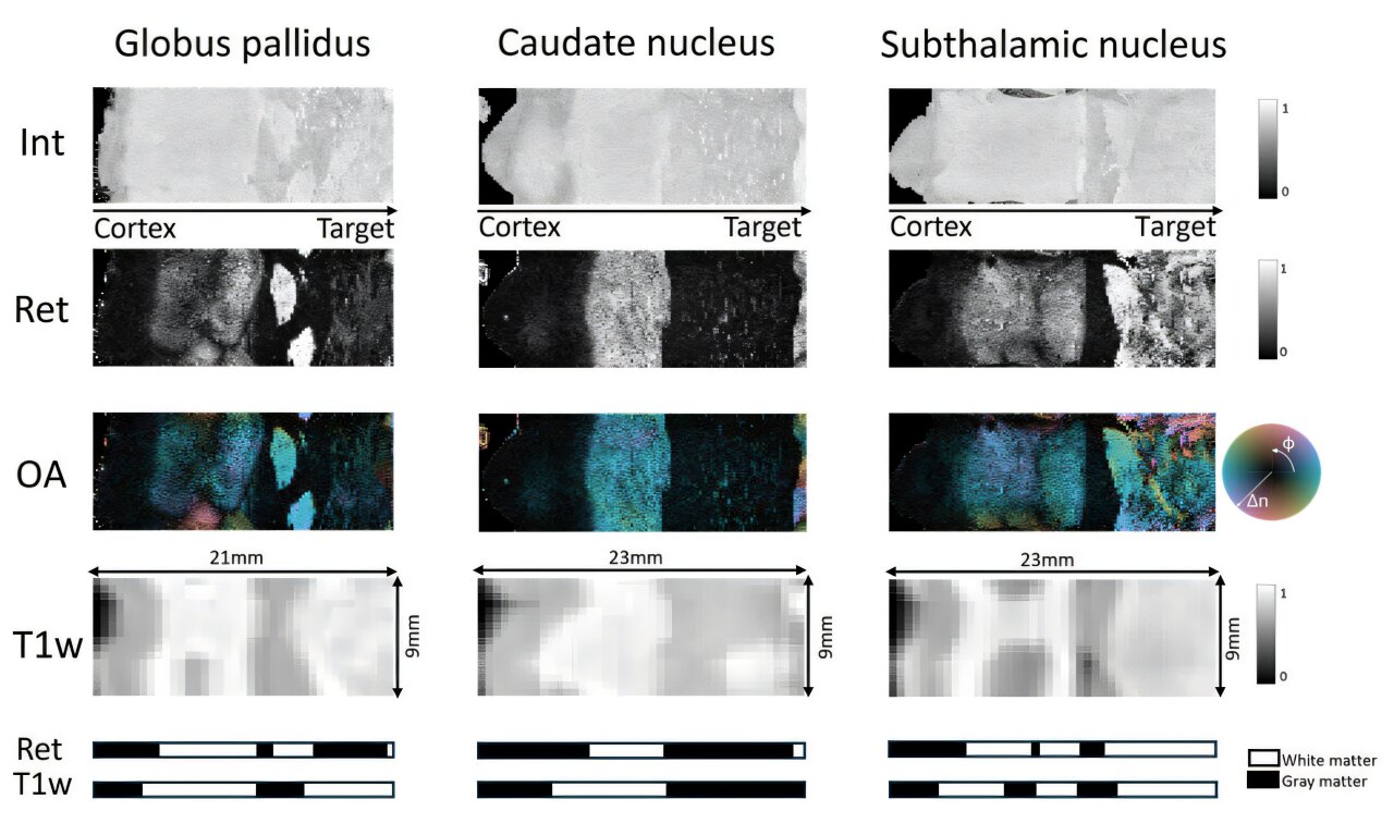
The results of the study showed that PS-OCT could distinguish between white and gray matter more clearly than MRI. It also revealed fine fiber structures that MRI missed, such as the internal capsule, a dense bundle of fibers crucial for DBS planning. In one case, PS-OCT identified highly organized fiber tracts near the globus pallidus externus (GPe) that were invisible in MRI scans. These findings suggest that PS-OCT could provide real-time feedback during DBS procedures, improving accuracy and reducing the risk of electrode misplacement.
To compare the two imaging methods, the team used a simplified segmentation approach. They averaged data along the probe path and applied clustering to separate tissue types. This allowed them to create "tissue barcodes" showing transitions between white and gray matter. PS-OCT produced sharper and more consistent barcodes than MRI, highlighting its potential for guiding electrode placement.
Despite its advantages, PS-OCT currently measures fiber orientation only in two dimensions. Future improvements could enable full 3D mapping, further enhancing its utility. Additionally, the PS-OCT probe used in the study was slightly larger than standard DBS electrodes, but smaller probes are already available and could be adapted for clinical use.
Shadi Masoumi, the corresponding author from Laval University, stated, "Catheter-based PS-OCT shows strong promise as a tool complementary to MRI in DBS neurosurgery. By providing high-resolution structural information and visualizing critical fiber pathways, it could help surgeons target brain regions more precisely."
Next steps for the research include live testing, integration into surgical workflows, and direct comparisons with diffusion MRI, another technique used to map brain fibers. If successful, PS-OCT could become a valuable addition to the neurosurgical toolkit, improving outcomes for patients undergoing DBS.
This groundbreaking study highlights the potential of advanced imaging techniques to revolutionize the field of neurosurgery, offering new hope for those suffering from neurological disorders.
Post a Comment for "Polarized Light Imaging Boosts Deep Brain Stimulation Accuracy"
Post a Comment