Cancer Cell Detection: Microfluidic Device Captures 90% of Cancer Cells in Blood
Understanding Circulating Tumor Cells and Their Detection Challenges
Circulating tumor cells (CTCs) are cancer cells that have detached from a primary tumor and entered the bloodstream. These cells can travel through the circulatory system and potentially form secondary tumors in other parts of the body. Detecting and analyzing CTCs from blood samples is crucial for early cancer diagnosis, treatment monitoring, and predicting disease progression. However, capturing these rare cells efficiently has proven to be a significant challenge in clinical settings.
The traditional methods of CTC detection often rely on antibodies to bind and isolate these cells. While effective, this approach requires complex chemical processes and can be costly when scaled up for widespread use. Researchers are continuously seeking more efficient and affordable alternatives to address these limitations.
Breakthrough in Microfluidic Technology
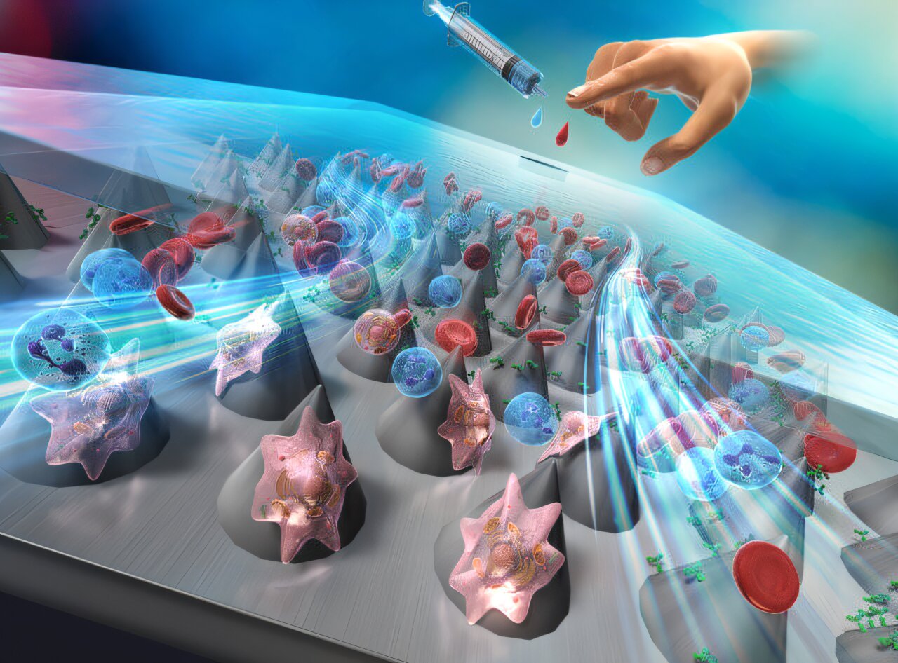
A recent breakthrough in microfluidic technology offers a promising solution. Scientists led by Professor Masumi Yamada from Chiba University in Japan have developed innovative microfluidic devices that incorporate microcones—structures comparable in size to biological cells. This new approach aims to enhance the efficiency of CTC capture while reducing costs.
The research team included Mr. Yuhei Saito from Chiba University and Dr. Shuhei Aoyama from Denka Innovation Center. Their findings were published in the journal Lab on a Chip on May 28, 2025. The study details how microcones, engineered with nanoscale roughness, can interact with antibodies to capture cancer cells effectively.
Design and Functionality of the Microfluidic Device
The researchers used polycarbonate (PC) sheets with microcone arrays created using thermal nanoimprint lithography (T-NIL), a heat-based fabrication technique. The microcones were approximately 30 micrometers in size and arranged in a hexagonal pattern. Their unique surface chemistry and morphology allowed them to bind and adsorb antibodies efficiently.
To create a functional microdevice, the scientists coated the PC sheets with anti-human epithelial cell adhesion molecule antibodies. These antibodies can specifically target and capture cancer cells. The coated PC sheets were then sandwiched between a flat plate and a glass slide to form microfluidic channels.
Optimizing Capture Efficiency
To understand how the microcone arrangement affects the capture of cancer cells, the researchers controlled the orientation angles of the microcone array within the microgap channels. Prof. Yamada explained, "We aimed to investigate how the microcone orientation impacts the capture behaviors of cancer cells."
During experiments, the microfluidic device demonstrated high selectivity in capturing human breast cancer (MCF-7) and lung cancer (A549) cells from blood samples. Notably, devices with microcone orientations at 15° or 30° maintained over 90% capture efficiency for MCF-7 cells even at high flow rates. This result highlights the importance of microcone arrangement in enhancing the performance of the microfluidic system.
Clinical Diagnostic Potential
To evaluate the diagnostic potential of the device, the researchers conducted immunostaining studies using fluorescent dyes. Despite using multiple reagents to label cells, they found that cancer cells remained trapped within the microchannels and did not escape. Under fluorescent light, the captured cancer cells could be easily distinguished from normal cells.
Prof. Yamada emphasized the significance of this development: "There are many technologies for detecting cancer, but it has been a long-standing challenge to detect cancer cells with high sensitivity using minimally invasive methods. We hope that through our new microfluidic system, even simple blood tests can be utilized to aid in the early diagnosis of cancer."
Future Implications
This study presents a novel, cost-effective, and highly sensitive diagnostic tool for detecting circulating cancer cells in blood. The microfluidic system offers a promising alternative to conventional methods, potentially revolutionizing cancer diagnostics and treatment monitoring. With further development and validation, this technology could become a vital asset in clinical settings, enabling earlier detection and improved patient outcomes.
Post a Comment for "Cancer Cell Detection: Microfluidic Device Captures 90% of Cancer Cells in Blood"
Post a Comment